ESPE2014 Poster Category 3 Sex Development (11 abstracts)
Gonadoblastoma and Papillary Tubal Hyperplasia in Ovotesticular Syndrome
Enver Simsek , Cigdem Binay , Baran Tokar , Sare Kabukcuoglu & Melek Ustun
Eskisehir Osmangazi University School of Medicine, Eskisehir, Turkey
Background: Ovotesticular disorder of sexual development (DSD is a rare form of DSD in which both testicular and ovarian tissues are present in the same individual either in a single gonad (ovotestis) or in opposite gonads with a testis and an ovary on each side.
Objective and hypotheses: To discuss rare cases of ovotesticular DSD and one of the novel findings of these cases.
Methods and patients: Case 1 is the first child of unrelated parents and was referred on the third day after birth due to ambigious genitalia. Upon physical examination, the patient had ambiguous genital including a phallus with a length of 2.3 cm, bifid labioscrotal folds, incomplete labioscrotal fusion, ventral opening of the urethra, chordea, and non-palpable gonads. Case 2 was a 15-year-old female presented with lack of pubertal development and primary amenorrhea. Physical examination revealed short stature (−2.1 SDS), Tanner stage I breast development, normal female external genitalia phenotype.
Results: Hormonal investigations of case 1 excluded congenital adrenal hyperplasia, leydig hypoplasia, 5α-reductase deficiency and androgen insensitivity syndrome. Chromosomal analysis and fluorescence in situ hybridisation of SRY revealed a SRY-positive 46,XX. Laparoscopic examination of case 1 revealed Mullerian remnants. Histopathological examination of bilateral gonadal biopsies showed ovotestes. Karyotype analysis and FISH of SRY of case 2 showed an SRY (+) 46,XY karyotype. Laparoscopic examination of case 2 revealed rudimentary Müllerian structures.
Conclusions: Laparoscopic examination and gonadal biopsy for histopathological diagnosis remain the cornerstones for a diagnosis of ovotesticular DSD.

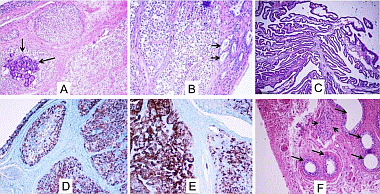
Figure 1 Histological examination of gonadoblastoma with superimposed dysgerminoma in the left gonad (A, B, C and D) and streak right gonad (E and F). (B) Sertoli cells (arrows) and dysgerminoma nests progressing to gonadoblastoma. The right streak gonad showed polypoid and papillary hyperplasia of the tubular epithelium. (F) Leydig cell remnants (arrowheads) and epididymis (arrows).
 }
}


