ESPE2014 Poster Presentations Adrenals & HP Axis (1) (11 abstracts)
A Novel Mutation in Exon 5 of TP53 Gene in a Familial Adrenocortical Carcinoma
Enver Simsek , Cigdem Binay , Baran Tokar , Emine Dündar & Meliha Demiral
Eskisehir Osmangazi University School of Medicine, Eskisehir, Turkey
Background: Adrenocortical carcinoma (ADCC) is a rare cancer in children and differs significantly in epidemiology, clinical characteristics, and biologic features from their counterparts in adults. Germline mutations of the TP53 tumor suppressor gene are associated with cancer predisposition in families with the Li-Fraumeni syndrome.
Aim: To report a young girl with family a history of ADCC who presented with severe virilisation secondary to ADCC, and to investigate the possibility the germline p53 tumour suppressor gene mutation in patient and the other family members.
Subjects and methods: In 2002, the first daughter was diagnosed with an ADCC at the age of 24-month-old. More than a decade later (2013), the second daughter presented with the symptoms and signs of severe virilisation at the age of 38-month-old and she was also diagnosed with an ADCC. Because of this family history, all of the family members underwent testing for a genetic mutation that is associated with increased cancer formation. Full sequencing of the coding exons (2 – 11) and associated splice junctions of the p53 gene was performed on patients and their healthy family member.
Results: Sequencing analysis showed a heterozygous GCACCCG deletion in exon 5 of TP53 gene (c.461_467delGCACCCG), which configures the mutation p.G154A.fs*14. This mutation has never been described in association with ADCC and was found in the proband and her affected sister. In addition, the same mutation was detected in two other siblings and father who have not yet developed any malignancy.
Conclusion: This observation of germline TP53 mutation in this family associated with ADCC confirms that genetic counselling and germline testing for TP53 should be offered to all pediatric patients with ADCC. These cases also highlight the need for close surveillance of patients and first degree relatives with TP53 gene mutation for malignancies

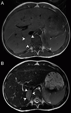
Figure 1 Right surrenal gland magnetic resonance imaging showing hypointense mass on T1 seguence (A), and hyperintense mass (2.0 X 2.5 cm) on T2 sequence after radiocontrast injection.
 }
}


